

ICU Topics > Mechanical Ventilation
Mechanical Ventilation
Mechanical ventilation is one of the most common interventions in the ICU — 85–90% of patients require it at some point during their stay. Most patients wean successfully once the underlying cause is resolved, but 20–30% require more time. Reintubation carries a 30–40% increase in mortality, which is why airway safety during therapy is not optional.
A patient can be connected to the ventilator in 1 of 3 ways:
Nasotracheal intubation (nose) – Short term
Endotracheal intubation [ETT] (mouth) – most common; short term
Tracheostomy (throat) – for extended vent weaning; long term
Translaryngeal tubes (ETT or nasotracheal) are measured to a specific depth. Tubing has hash marks with numbers to indicate the distance until the end of the tubing. *Tubing should not go into/out of the mouth/throat further. Take note of the position of the tubing and measurement at the lips or nostrils. Monitor before/during/after transfers.
A cuff is inflated around the tube to ensure that the air is being delivered directly into the lungs. If a cuff leak is suspected, the patient may be able to phonate or make audible sounds from the mouth. Alert RN immediately.
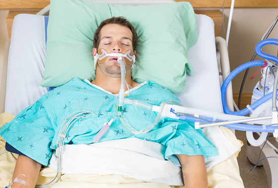
Endotracheal Intubation (ETT)
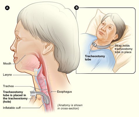
Tracheostomy
Video Overview
Before You Treat: What to Check
Before initiating therapy with a mechanically ventilated patient, review the following on the ventilator screen and in the chart:
Mode: What level of support is the patient receiving? Volume/Pressure Control indicates full vent dependence. SIMV and PSV indicate active weaning.
Rate: What is the set respiratory rate? On A/C mode, the rate is preset — the patient cannot increase their RR to compensate during activity.
FiO2: Is the patient requiring high FiO2 (>60%)? High oxygen needs suggest limited reserve — consider deferring or significantly modifying the session.
PEEP: PEEP >15 cm H2O indicates severe lung injury. Mobility is not indicated at this level.
Pressure Support: PS ≥20 cm H2O indicates high vent dependence and likely not appropriate for mobility.
SpO2 trend: Is the patient maintaining adequate oxygenation at rest? Is it stable or fluctuating?
Vitals trend: Is the patient hemodynamically stable? Review recent HR, BP, and RR trends before entering the room.
Sedation: What medications are running? Can the dose be safely reduced for therapy? Review the medications table below.
Orders: Confirm the patient has a therapy order and check for specific parameters or precautions.
Communicate: Check in with nursing and RT before starting, particularly for higher-acuity patients. RT can tell you whether the patient is currently on a weaning trial or has an SBT planned.
Terms and Norms
Tidal Volume (VT): amount of air inhaled or exhaled with each breath (based on weight).
Average healthy male adult: 500 ml.
Positive End Expiratory Pressure (PEEP): pressure applied by the vent at the end of each breath. Varies and can be increased or decreased as needed. There is a constant amount of baseline positive airway pressure (i.e. 5 cm H20) that’s maintained even at end of expiration. Assists in keeping alveoli open to minimize atelectasis.
3-5 cm H2O (low): physiologic; used to preserve normal functional residual capacity (FRC=expiratory receiver volume + residual volume)
5-15 cm H2O: used to treat refractory hypoxemia in acute lung injury.
Therapy may be appropriate; monitor SpO2 and respiratory status closely during activity
>15 cm H2O (high): used only for severe lung injury (likely candidates for proning or ECMO). Mobility not indicated.
Pressure Support (PS) norms: 0 cmH2O to 30 cmH2O. Usually 5-15 cmH2O.
≤ 5 cmH20 LOW vent settings; may be ready to extubate.
≥ 20 cmH20 HIGH vent settings; Not stable/likely not ready for mobility.
FiO2 (fraction of inspired oxygen): concentration of oxygen in the gas mixture.
“Room air” is 21% FiO2 (or 0.21). Usually see 30-60% (0.3-0.6) for a patient requiring vent support.
Vent can provide up to 100% FiO2. Patient who requires high FiO2 support likely unstable/not appropriate for therapy; If Patient decompensates, additional support is limited.
Spontaneous Breathing Trial (SBT)
A spontaneous breathing trial is a test of the patient's ability to breathe independently. The patient is placed on minimal ventilator support — typically low-level pressure support or a T-piece — for a set period to assess readiness for extubation.
Do not schedule therapy during an SBT. The patient is already under respiratory stress from the trial itself, and adding the demands of activity can cause trial failure or patient decompensation. Before starting any session, confirm with nursing or RT whether an SBT is planned or currently in progress.
Ventilator Modes & Settings
Determined by the MD and RT. Settings will vary depending on the patient’s needs.
Control mode is the most dependent
Patients generally progress from more dependent to less dependent modes prior to extubation. You might see a progression from synchronized intermittent mandatory ventilation (SIMV) to pressure support volume (PSV) and to continuous positive airway pressure (CPAP) prior to the ability to be extubated.
Modes of Ventilation
| Mode | Type of Support | Description | Therapy Implications |
|---|---|---|---|
| Volume Control | Total assist | Preset volume of air is delivered with each breath for lung inflation. | Patient is sedated and likely paralyzed. Limit activity to gentle PROM. |
| Pressure Control | Total assist | Air is delivered until a preset pressure is reached in the lungs. | See Volume Control above. |
| Assist-Control (A/C) | Partial assist; often used with sedated patients | Volume cycled with a preset number of breaths. Patient can initiate each mechanical breath but cannot take spontaneous unassisted breaths between mechanical breaths. | Minimal work of breathing. If not in sync with the vent, patient may "buck" or fight the vent. If able to participate, patient can become short of breath during activity — breathing rate is preset, so the patient cannot increase RR to compensate. Monitor vitals and signs of anxiety. |
| Synchronized Intermittent Mandatory Ventilation (SIMV) | Weaning mode | Volume cycled with a preset number of mandatory breaths. Allows spontaneous unassisted breathing between mechanical breaths. Used as respiratory exercise during the weaning process. | Increased work of breathing. Respiratory muscle fatigue is common. Schedule therapy after weaning trials to avoid overtiring the patient. |
| Pressure Support (PSV) | Weaning mode; spontaneous breathing required | Minimum pressure support that compensates for the resistance of the vent tubing. Increased pressure may be provided to reduce work of breathing. | Increased work of breathing. Schedule therapy during weaning trials. The team may want an assessment of the patient's ability to perform activity on a lower vent setting. |
Note. Adapted from "Intensive Care Unit," by K. Clark, 2017, Table 9.6, p. 120, in Occupational Therapy in Acute Care. AOTA Press. Copyright 2017 by AOTA Press. Reprinted with permission.
Most Common Ventilator Modes
-
Assist-Control (A/C) (Respiratory Therapy Zone, 2021)
Non-weaning mode. Pre-set: Rate & Vt
Minimum number of pre-set mandatory breaths are delivered by the vent, Patient CAN trigger assisted breaths. However, the vent will deliver pre-set Vt when this happens. Doesn’t allow for spontaneous breaths.
Can be volume (more common) or pressure-assist control.
On Hamilton G-5 vent mode will read: (S)CMV for volume A/C or P-CMV for pressure A/C
Parameters that should be noted ON the ventilator: Confirm: mode, rate, target variable (volume OR pressure control), FiO2, PEEP

-
Synchronized Intermittent Mandatory Ventilation (SIMV) (Respiratory Therapy Zone, 2021)
Weaning mode. Pre-Set: Rate & Vt
Vent delivers a pre-set minimum number of mandatory breaths. Patient CAN breathe spontaneously between vent breaths.
Each spontaneous breath receives a Vt dependent on the Patient’s effort NOT the pre-set Vt. **Often used with PSV to help overcome the resistance of vent tubing on spontaneous breaths.
On Hamilton G-5 vent mode will read: SIMV
Parameters that should be noted ON the ventilator: Confirm: mode, rate, target variable (Volume OR Pressure control), FiO2, PEEP, Pressure support

-
Pressure Support Ventilation (PSV) (Respiratory Therapy Zone, 2021)
Spontaneous breaths supported by the vent during inspiratory phase of breathing. When Patient triggers a breath, vent assists by adding pressure to make breathing easier. May be applied to spontaneous breathing during SIMV or CPAP.
Once the patient triggers the vent: Pre-set positive pressure is delivered. Volume is NOT pre-set. PS augments Vt.
Patient in control of RR & inspiratory time.
No guaranteed ventilation or RR. (Except back up apnea rate).
On Hamilton G-5 vent mode will read: SPONT
Parameters that should be noted ON the ventilator: Confirm: mode, PS, FiO2, and PEEP

-
CPAP
When used as a ventilator mode, CPAP delivers continuous positive airway pressure throughout the breathing cycle via the ETT or tracheostomy — unlike non-invasive CPAP delivered through a face mask. The vent provides no mandatory breaths and no pressure support. The patient breathes entirely on their own.
CPAP mode is typically a late weaning step and indicates the patient is close to extubation.
On Hamilton G-5 vent mode will read: CPAP
Parameters to confirm on the ventilator: mode, FiO2, PEEP
Therapy implications: The patient is performing the full work of breathing — this is a positive sign clinically. However, monitor closely for respiratory fatigue during activity. Coordinate session timing with the weaning team and consider starting with lower-intensity tasks.
Common Medications while Mechanically Ventilated
| Drug | Mechanism | Dosing (drip) | Onset | Half-Life | Therapy Implications |
|---|---|---|---|---|---|
| Propofol (Diprivan) |
Short-acting CNS depressant; decreases level of consciousness | 5–50 mcg/kg/min | Seconds | 3–12 hrs | Consider dose and ability to follow commands. Can the patient tolerate a lower dose? A decrease should have a quick impact on arousal. |
| Dexmedetomidine (Precedex) |
Provides sedation without high risk of respiratory depression; allows semi-arousal | 0.2–0.7 mcg/kg/hr | 1 min | 2 hrs | Sleep/wake cycle preserved. Patient is usually more awake compared to other sedatives. |
| Midazolam (Versed) |
CNS depressant; causes relaxation and amnesia without general anesthesia | 1–30 mg/hr | 1–5 min | ~3 hrs | Consider dose and ability to follow commands. Can the patient tolerate a lower dose? |
| Fentanyl (Sublimaze) |
Synthetic opioid; reduces perception of pain with sedative effects | 1–2 mcg/kg IV bolus or 1–2 mcg/kg/hr infusion | 7–8 min | 3–4 hrs | Bolus or continuous infusion? Is pain a limiting factor for participation? |
| Cisatracurium (Nimbex) |
Skeletal muscle relaxant; blocks acetylcholine at the neuromuscular junction causing chemical paralysis | 1–10 mcg/kg/min | 2–3 min | 22 min | Patient is chemically paralyzed. OT/PT is not indicated. |
Safety
Note: vent tube disconnection is common and very different from accidental extubation — know the difference before you're in the room.
Accidental Extubation Protocol:
Prepare: be familiar with the ambu bag’s location in the room and have a plan of action in mind
Prevent: do not underestimate the patient’s determination to self-extubate. Keep your eyes on the patient at all times and be prepared that they might try to pull out the tubing.
Follow a plan: 1) Call for the RN and place the patient in a safe position; 2) while maintaining the patient’s physical safety, retrieve the ambu bag, 3) begin providing rescue breaths and attach the ambu bag to oxygen
(Clark, 2017)
Common Reasons for Intubation in the ICU
(Howard et al., 2003)
Generally required for airway management with intubation indicated for patients with:
Impaired level of consciousness (GCS <9)
Progressive respiratory impairment/failure
Inability to oxygenate (due to high secretions) or ventilate (i.e., myasthenia gravis)
Impaired cough or airway clearance
Pulmonary edema/aspiration
Seizure activity
Further Reading
Miller, N. (2013). Set the stage for ventilator settings. Nursing Made Incredibly Easy, 11(3), 44–52. https://doi.org/10.1097/01.NME.0000428429.60123.f7
Zanni, J. (n.d.). Understanding Mechanical Ventilation. https://www.johnshopkinssolutions.com/wp-content/uploads/2017/10/4-Understanding-Mechanical-Ventilation.pdf
References
Clark, K. (2017). Intensive Care Unit. In H. Smith-Gabai & S. E. Holm (Eds.), Occupational Therapy in Acute Care (2nd ed., pp. 115–135). AOTA Press.
Howard, R. S., Kullmann, D. M., & Hirsch, N. P. (2003). Admission to neurological intensive care: Who, when, and why? Journal of Neurology, Neurosurgery & Psychiatry, 74(suppl 3), iii2–iii9. https://doi.org/10.1136/jnnp.74.suppl_3.iii2
Respiratory Therapy Zone. (2021). Ventilator modes made easy (study guide for mechanical ventilation). Respiratory Therapy Zone. www.respiratorytherapyzone.com/ventilator-modes-practice-questions/
Zanni, J. (n.d.). Understanding Mechanical Ventilation. https://www.johnshopkinssolutions.com/wp-content/uploads/2017/10/4-Understanding-Mechanical-Ventilation.pdf